The analysis of dissolved inorganic carbon in liquid using a microfluidic conductivity sensor with membrane separation of CO2.

The analysis of dissolved inorganic carbon in liquid using a microfluidic conductivity sensor with membrane separation of CO2.

Autonomous regular analysis of oceanic dissolved inorganic carbon (DIC) focus with depth is of good significance with regard to ocean acidification and native climate change.

However, miniaturisation of in situ analysis applications is hampered by the size, worth and vitality requirements of standard optical instrumentation.

Here, we report a low-cost microfluidic totally different based totally on CO2 separation and conductance measurements that might consequence in built-in lab-on-chip applications for ocean float deployment, or for moored or autonomous flooring automotive functions.

Conductimetric dedication of focus, in the seawater differ of 1000-3000 µmol kg-1, has been achieved using a microfluidic thin-film electrode conductivity cell and a membrane-based gas alternate cell. Sample acidification launched CO2 by manner of the membrane, reacting in a NaOH service, later drawn by manner of a sub-µL conductivity cell, for impedance versus time measurements. Precision values (relative commonplace deviations) had been ~ 0.2% for peak peak measurements at 2000 µmol kg-1.

Comparable precision values of ~ 0.25% had been obtained using a C4D electrophoresis headstage with comparable measurement amount. The required full sample and reagent volumes had been ~ 500 µL for the low amount planar membrane gas alternate cell. In distinction, earlier conductivity-based DIC analysis applications required full volumes between 5000 and 10,000 µL.

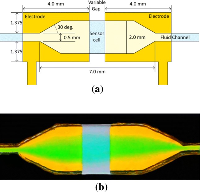
Long membrane tubes and macroscopic wire electrodes had been averted by incorporating a planar membrane (PDMS) in the gas alternate cell, and by sputter deposition of Ti/Au electrodes immediately onto a thermoplastic (PMMA) manifold.

Future effectivity enhancements will sort out membrane chemical and mechanical stability, extra amount low cost, and ingredient integration into a single manifold.

The analysis of dissolved inorganic carbon in liquid using a microfluidic conductivity sensor with membrane separation of CO2.

Overcoming the bottleneck to widespread testing: A speedy consider of nucleic acid testing approaches for COVID-19 detection.

The current COVID-19 pandemic presents a vital public effectively being catastrophe, and a larger understanding of the scope and unfold of the virus may very well be aided by additional widespread testing. Nucleic-acid based totally exams on the second provide basically essentially the most delicate and early detection of COVID-19.

However, the “gold commonplace” take a have a look at pioneered by the United States Center for Disease Control & Prevention, takes a quantity of hours to complete and requires in depth human labor, provides much like RNA extraction kits that might develop into in temporary present and relatively scarce qPCR machines.

It is clear that a monumental effort should be made to scale up current COVID-19 testing by orders of magnitude. There is thus a pressing need to guage totally different protocols, reagents, and approaches to allow nucleic-acid testing to proceed in the face of these potential shortages. There has been a giant explosion in the amount of papers written all through the primary weeks of the pandemic evaluating potential advances, comparable reagents, and choices to the “gold-standard” CDC RT-PCR take a have a look at.

Here we present a assortment of these present advances in COVID-19 nucleic acid testing, collectively with every peer-reviewed and preprint articles.

Due to the speedy developments all through this catastrophe, we have included as many publications as doable, nevertheless many of the cited sources have not however been peer-reviewed, so we urge researchers to extra validate outcomes in their very personal labs.

We hope that this consider can urgently consolidate and disseminate information to help researchers in designing and implementing optimized COVID-19 testing protocols to increase the provision, accuracy, and tempo of widespread COVID-19 testing.

NMR quantification of trimethylamine-N-oxide in human serum and plasma in the clinical laboratory setting.

NMR quantification of trimethylamine-N-oxide in human serum and plasma in the clinical laboratory setting.

OBJECTIVETrimethylamine-N-oxide (TMAO) produced by intestine microbiota metabolism of dietary choline and carnitine has been proven to be related to elevated threat of heart problems (CVD) and to offer incremental clinical prognostic utility past conventional threat elements for assessing a affected person’s CVD threat.

The purpose of this research was to develop an automatic nuclear magnetic resonance (NMR) spectroscopy assay for quantification of TMAO focus in serum and plasma utilizing a high-throughput NMR clinical analyzer.

METHODSKey steps in assay improvement included:

(i) shifting the TMAO analyte peak to a much less crowded area of the spectrum with a pH buffer/reagent, (ii) attenuating the broad protein background sign in the spectrum and

(iii) utilizing a non-negative least squares algorithm for peak deconvolution. Assay efficiency was evaluated in keeping with Clinical and Laboratory Standards Institute pointers.

A way comparability research was carried out to check TMAO concentrations quantified by NMR and mass spectrometry (MS).RESULTSThe within-run and within-lab imprecision ranged from 4.Three to 14.5%. Under the acquisition technique employed, the NMR assay had a restrict of clean, detection and quantitation of 1.6, 3.0 and 3.3μM, respectively. Linearity was demonstrated inside the reportable vary of 3.Three to 3000μM.

TMAO measurements utilizing the NMR assay, which entails minimal pattern preparation, in contrast effectively with values obtained with the MS-based assay (R2=0.98).CONCLUSIONSThe NMR primarily based assay offers a easy and correct measurement of circulating TMAO ranges amenable to the high-throughput calls for of the clinical chemistry laboratory.

Moreover, assay efficiency allows the ranges of TMAO to be quantified in serum or plasma at clinically actionable concentrations for the evaluation of heart problems dangers and individualized dietary monitoring.

NMR quantification of trimethylamine-N-oxide in human serum and plasma in the clinical laboratory setting.

MAPS-seq: magnetic bead-assisted parallel single-cell gene expression profiling.

Recently developed single-cell RNA sequencing strategies enable the simultaneous profiling of the transcriptomes of hundreds of particular person cells.

However, present strategies nonetheless require superior tools or entail substantial waste of reagents. Here, we introduce magnetic bead-assisted parallel single-cell gene expression sequencing (MAPS-seq), a microwell-based technique that swimming pools samples earlier than the reverse transcription step, rising the ease of pattern preparation and lowering reagent waste.

Moreover, as a result of this technique makes use of common reagents and normal molecular biology lab devices, it’s straightforward to implement, even in labs that haven’t beforehand carried out single-cell RNA sequencing.

We validated our technique by demonstrating that it may well generate gene expression information at the single-cell stage. We then utilized the MAPS-seq technique to investigate 237 human myelogenous leukemia cells handled with one of three totally different medication or dimethyl sulfoxide.

We noticed transcriptional modifications and recognized marker genes that point out a drug response. Furthermore, the MAPS-seq technique produced information of comparable high quality to these of present single-cell RNA sequencing strategies.

Consequently, we anticipate that our technique will present researchers with a extra accessible, much less wasteful, and much less burdensome technique for investigating the transcriptomes of particular person cells.

Adenovirus infections:

Adenovirus
Adenovirus

What are the Adenovirus infections?

Adenoviruses are DNA viruses that are classified according to the presence of 3 major antigens in the capsid (hexone, pentone, and fiber). There are 7 species of human adenoviruses (A to G) and 57 serotypes. Different serotypes are associated with different diseases.

In general, the infection is contracted by contact with secretions (including on the fingers of infected patients) from an infected person or with a contaminated object (eg, towels, instruments). The infection can be transmitted by air or water (eg, by swimming in lakes or swimming pools without adequate chlorine). Asymptomatic respiratory or gastrointestinal viral desquamation can continue for several months or even years.

Signs and symptoms

In immunocompetent hosts, most adenovirus infections are asymptomatic. When infections are symptomatic, a broad spectrum of clinical manifestations can occur because most adenoviruses that cause mild disease have an affinity for a variety of tissues.


Most symptomatic infections occur in children and cause fever and upper respiratory symptoms, such as pharyngitis, otitis media, cough, and exudative tonsillitis with cervical lymphadenopathy, which may be difficult to distinguish from group A strep throat. Adenoviruses Types 3 and 7 cause a syndrome characterized by conjunctivitis, pharyngitis, and fever (pharyngoconjunctival fever).


Very rarely, some adenovirus syndromes, found in infants, can manifest with severe bronchiolitis and pneumonia. In closed populations of young adults (eg military recruits), outbreaks of respiratory illness, with fever and symptoms of the lower airways, usually tracheobronchitis, may occur, but also occasionally pneumonia.


Clusters of cases of severe respiratory diseases caused by specific adenoviruses (particularly types 7, 14, and 55) have occurred in healthy adults. Adenovirus infections are increasingly recognized as the cause of severe respiratory diseases and other clinical diseases in immunocompromised adults.


Epidemic keratoconjunctivitis is sometimes severe and, sporadically, can cause epidemics. Conjunctivitis is usually bilateral and preauricular lymphadenopathy is often palpable. Chemosis, pain, and visible punctate corneal lesions can also be identified with fluorescein staining. Systemic signs and symptoms are mild or absent. Epidemic keratoconjunctivitis usually resolves in 3 to 4 weeks, although corneal lesions can persist much longer.


Non-respiratory adenoviral syndromes include hemorrhagic cystitis, diarrhea in infants, and meningoencephalitis.

Most of the patients recover completely. Even severe primary adenovirus pneumonia is not fatal, except in rare fulminant cases, especially in infants, military recruits, and immunocompromised patients.

Clinical evaluation


For severe disease, polymerase chain reaction (PCR) tests on respiratory secretions and blood.
Laboratory diagnosis of adenovirus rarely affects management. During acute illness, the virus can be isolated from respiratory and eye secretions and is often found in faeces and urine. A 4-fold increase in serum titer indicates recent adenovirus infection.

PCR tests can detect adenovirus DNA in respiratory secretions and blood and are useful when patients have severe disease and a diagnosis is needed.

Why utilize adenovirus?

  • High disease efficiency and subsequent performance of recombinant protein.
  • Broad assortment of hosts (divided and undivided, stem cells and primary tissues ).
  • Non-integrative (without danger of affecting the expression of this host receptor ).
  • Low immunogenicity (post-infection cell viability is extremely large ).
  • It may be utilized in vivo and in vitro.
  • Biosecurity: We utilize replication incompetent human adenoviruses (-E1 / -E3) type 5 (Ad5).

General Facilities

Intellectual Property

Because the consequence of an extremely effective SBIR application, KAIROS retains an abundance of intellectual property which transcend historical dates of conception and supports pending patents. Awards and contracts include:


Enzyme Evolution

KAIROS has generated new technologies in solid phase receptor screening which combines innovative imaging apparatus with directed development. We provide your enzyme’s action to enhance, or we could put a tool on your website.
General Facilities • KAIROS has a rather active genetic engineering team that’s focusing on proteins that are intrinsically fluorescent and about the directed evolution of enzymes. Biochemistry our cell biology, and molecular biology classes operate within precisely exactly the environment which fosters the co-development of both applications and instrumentation.

Macromolecular Scaffolds for Energy Research (DOE)
Quantitative FRET Microscopy (NIH)

Chromogenic Winogradsky Movies for Enzyme Discovery (NIH)
Directed Evolution of Enzymes With Optical Sorting (NIH)
Imaging Facilities •

KAIROS is ongoing to create both model and turn-key electronic imaging spectrophotometers (DIS) effective at specifying the fluorescence excitation, fluorescence emission, and absorption spectra for each and each single pixel or set of pixels at a field of opinion. All programs have been calibrated. We construct and design imaging spectrophotometers that are personalized to match requirements and the demands of researchers. Instruments various prototype programs under development along with MicroDIS MicroFRET Kcat.
By incorporating optical design and applications technology using molecular genetics, KAIROS’ multidisciplinary research is targeted toward creating novel instrumentation, reagents, and methods from the fields of biotechnology, microscopy, medication, and materials science.
Business Information •

Considering our very first imaging spectrophotometer, we’ve enlarged our cognitive engineering and cell research amenities. KAIROS has been the very first organization permit and to develop a specially GFP derivative.

  • Visualizing Post-Translational Modifications (NIH)Multispectral
  • Bacterial Identification (NIH)
  • Strong Phase 2 Screening (NIH)
  • Optimization of Cellular Screen (NIH)